|
The following of photographs show Sex Re-assignment Surgery (SRS) being performed on a male-to-female patient using a variant of the penile inversion procedure. The photos are taken from a lecture video put in the public domain by the University of California, San Francisco, USA. The video uses the term "Gender Affirmation Surgery", and the procedure was performed by Dr Maurice Garcia. Additional feminisation surgery not evident in the selected photos was performed by Dr Thomas Satterwhite. As reference to help understand the photo's, it's useful to compare diagrammes of the male and female reproductive systems:
The human male and female reproductive systems have the same structural origins. For example, the male gonads, or sex glands (the testes) and the female gonads (the ovaries) derive from the same rudimentary sex glands in the foetus. Similarly, the clitoris in the female corresponds to the penis in the male. However important organs such the female uterus or the male prostate never develop in the other sex. The actual appearance (external and internal) of the male and female 'bottom' is very different, with major surgery required to make either an approximate and infertile version of the other.
Photo 1. (Below) The patients penile and scrotal skin is prepared for removal, this will later be used to line the new vagina (neovagina). . Photo 2. Creation of a vaginal cavity.
Photo 3. Removal of the testes.
Photo 4. The urethra tube used for “peeing” will no longer pass though a penis and is shortened from about 20 cm to a 4 cm stump.
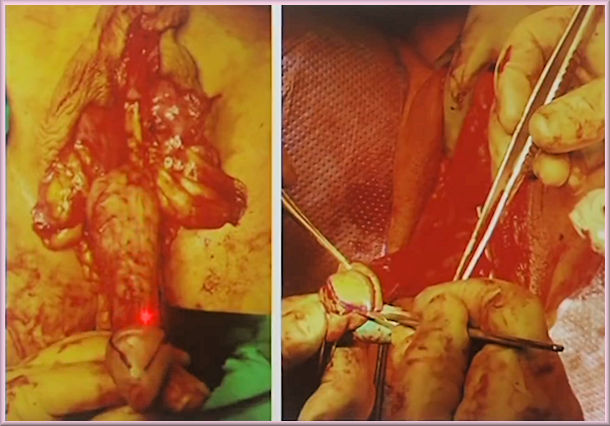
Photo 5. Clitoroplasty – the tip of the penis is removed and used to create a clitoris.
Photo 6. The neurovascular bundles (nerves and veins) leading to the glan penis are separated from the now surplus penile erectile tissue.
Photo 7. The remains of the penis are removed and the base of its erectile tissue (the corporal stump) sewn up.
Photo 8. The clitoris is added (sutured) at the top of the corporal stump and nerve bundles, just below is the still prominent urethal stump, which in turn is above the vaginal opening.
Photo 9. Women have a very different urinary system from men. The urethral stump is divided in to a pipe and spongy tissue. The pipe for urine (the urethral lumen) is positioned to avoid the vagina and exits the vulva directly just below the clitoris, the spongy tissue that protected the pipe when in the penis is used to form the urethral plate.
Photo 10. A tube covered by penile and scrotal skin is prepared and inserted in to the neovagina to line it.
Photo 11. The lining of the neovagina is over sewn. The relative positions of the clitoris and urethra above the vaginal opening is checked and adjusted.
Photo 12. Cleaned up appearance on the table.
Photo 13. The creation of a protective hood for the clitoris. Note: This procedure is often performed a few months later when the vulva has largely healed and a much better result is likely, it's also an opportunity to "tidy up" the external appearance of the area.
Photo 14. Packing of the neovagina and dressing of the surgery area.
Photo 15. Appearance of the patient's vulva area three and five months after surgery.
Photo 16. A final photo taken six months after SRS. Although the photo is of poor quality, the external result seems [unsurprisingly] to be very good. Scars are still obvious left and right of the vulva - but these are unavoidable.
|