|
[This
article should be read in conjunction with the article on Average
Body Size.
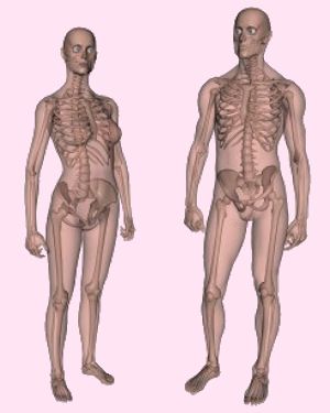
One of the distinctions between a genetic man and a genetic woman is the characteristics of their skeleton. The skeleton obviously sets or heavily influences the body size and its proportions. It's thought that genes (usually female "XX" or male "XY") mainly determine the basic size and shape of the skeleton, however some differences are exaggerated or emphasised at puberty by the sex hormones that surge around the growing body. The high levels of testosterone that appear in boys at puberty help lengthen and rugged'ise their still developing bones, enhancing and developing male skeletal characteristics such greater height and narrower pelvic width. The near absence in girls of these hormones prevents such skeletal developments; indeed the presence of high levels of oestrogen in a pubertal girl probably helps stimulate the growth and shape of her pelvic bones, but otherwise actually act to limit bone growth and final adult height. [Appropriate hormonal treatment in a young transsexual can thus have substantial benefits in terms of skeletal development, benefits not obtainable after puberty when bone development is essentially complete]. Despite the sex related differences, overall the differences between the skeletons of male and female bodies are actually surprisingly small compared with the similarities, as is illustrated by comparing the following diagrams:
Skeletal Sex Differentiation
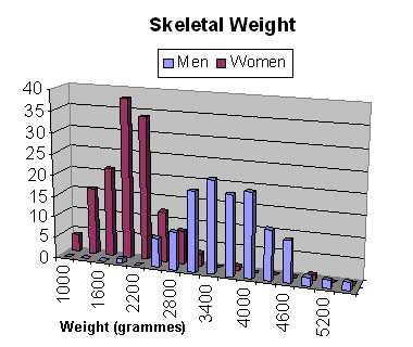
On average a male skeleton is larger than the female skeleton so this is a differentiation, but there is also a considerable overlap in skeletal size between the sexes - e.g. there are short men and tall women - so this is hardly a reliable method on its own. Other significant differences between male and female skeletons are that female bones are usually lighter and thinner than more robust male bones; female head bones are smaller and more lightly built; and the female pelvis is shallower and wider than the male's. This latter difference makes childbirth easier. The pelvis is thus considered to be the best area to determine or estimate the sex of a skeleton, while the skull (cranium and mandible) and postcranial skeleton is the second best area.
Skull and bone features vary from male to female - differentiation is usually based on the generalization that "typical male" features are more pronounced and marked than the same features in a female. By observing all the possible differentiating features of skeleton in a cumulative manner, it's possible to correctly identify the sex of a Caucasoid skeleton in about 90% of cases. Krogman ranks accuracy of sex determination using the pelvis at 95%, followed by the skull at 92%, the mandible alone at 90%, and long bone measures at 80%. Stewart indicates slightly lower yields, however the order of accuracy was the same, with the long bones again the least accurate. Success rates are somewhat lower for Negroid and Mongoloid skeletons. Note that it is often impossible to place absolute metric value on what constitutes a male feature, and what constitutes a female. Krogman addressed the difficulty of sex determination from skeletons when he stated: "here is the problem of subjectivity versus objectivity, of description versus measurement, of ‘experience’ versus statistical ‘standardization.’ "
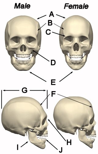
Skull Major differences between the female and male skill include the posterior of the cranium (the occipital), robusticity of the browridge, mastoid process, nuchal crest, temporal lines, and mandible. Although distinct, the ability to quantify measures of the skull for sex determination has met with limited success and successful sex identification based purely on a skull is a very subjective process based experience in identifying and assessing non-metric characteristics.
Above the orbits (eye sockets), the male cranium tends to have "blunt" superior margins and larger supraorbital (brow) ridges. The female cranium tends to have "sharp" superior margins of the orbits and no discernable supraorbital ridges.
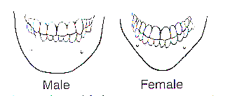
The mandible of a female cranium tends to have a "pointed" chin. The area around the gonial angle is smooth and does not project. The male mandible tends to have a "square" shape and in extreme case the area around the gonial angle is "flared". The dentition (teeth) of males is frequently larger. Finally,
the area of the temporal in the female cranium is smoother and less rugged
than that of the male cranium. In the female cranium the zygomatic
arch normally does not extend, as a ridge, posterior of the external
auditory meatus. In male crania the zygomatic arch typically
extends, as a ridge, posterior to the external auditory meatus. In
females the mastoid process is small and smooth. In males the
mastoid process is large and rugged.
Here is a summary of the differential criteria between the male and female cranium bones:
*These may vary. Exceptions occur frequently.
An ever increasing number of teenagers are having puberty blockers followed by "gender confirmation" hormone therapy . The result is a reduction in the differentiation between male and female skeletons. This will become statistically significant if 1% young teenagers identify as being transgender, which is very probable based upon recent trends.
However, as ever, there is great variance - many handsome actors on close examination have some feminine facial characteristics, while many supermodels have some very male characteristics. In absolute measures almost all dimensions of the female skull and face are smaller compared to the male features. The facial width is relatively larger in women than in men. Resulting contours are therefore more rounded in females, especially in the orbital area, with more prominent malar (cheek) bones and less prominent mandibular (chin/jaw) angles.
Forehead
Nose
Chin
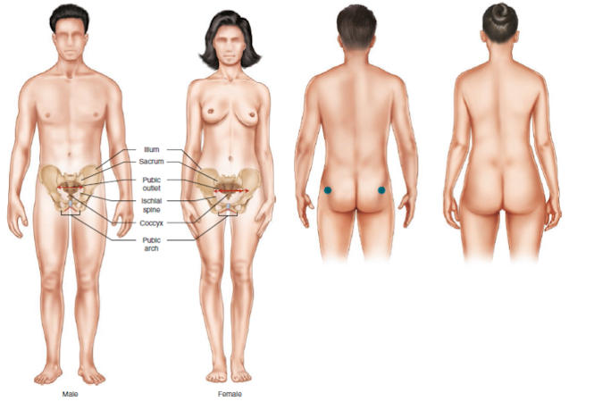
Another graphic showing the differences in female and male boney structures, and the resulting physical appearance, particularly in the pelvic region where differentition is greatest. Women generally having more obtuse subpubic angles, wider pelvic brims, and flared iliac crests. Viewed from the rear, young women also tend to have far more tissue (mostly fat) in their gluteal region (i.e. buttocks). with a distinct transition from a slimmer lower back, i.e. a waistline. By comparison, male buttocks tend to be flat or even have a distinct concavity, with a less obvious transition from the waist.
The pelvic girdle is formed by the sacrum, coccyx, and the two coxae. A coxa is formed by the fusion of three bones, the ilium, ischium, and pubis, which meet in the acetabulum or hip socket. At the back each coxa is attached by strong ligaments to the sacrum (base of spinal cord), and in front to each other at the pubic symphysis joint. This joint allows only slight bending movement, but it softens and becomes more flexible in a female giving birth. [Note: Other names for the pelvic bone are innominate bone and coxal bone.] Factors contributing to the overall shape of the pelvis are constrained by both the demands of bipedal locomotion, as well as those for perpetuating the human species - principally in relation to the requirements of childbirth. It is usually impossible to distinguish between the pelvis of a boy and a girl before puberty. Thereafter - of all bones - the pelvis shows the greatest sexual differentiation and is thus a primary tool for identifying male vs female skeletons post-puberty.
On average, the adult male pelvis is much heavier and narrower than that of the female. In comparison the female pelvis is broad and shallow, the geometry is designed with a greater outlet for passage through its bony openings of a baby's head and shoulders during birth. The female pelvis is also less massive and more delicate and its muscular impressions are slightly marked. A triangular shaped pubis with a broad medial aspect and no evidence of a ventral arc is a characteristically male pattern. The female pattern for these features is a rectangular pubis, pronounced ventral arc, and sharp, narrow medial aspect of the ischiopubic ramus. According to Bass, the presence of a ventral arc is the most diagnostic of the female pubic features.
In the female pelvis the ilia are less sloped, and the anterior iliac spines more widely separated; hence the greater lateral prominence of the hips. The pelvic inlet of females is larger and has a greater absolute circumference. The body of the pubis is longer, thereby increasing the size of the pelvic outlet. These characteristics all reach a maximum in a woman's mid- to late-20's - just before her fertility starts to decline significantly. The size of the pelvis varies not only in the two sexes, but also in different members of the same sex, and does not appear to be influenced in any way by the height of the individual. As a result, women of a short stature tend to have obviously very broad hips.
Sacrum Sacral
Index
Comparison of the Males and Female Pelvises:
Sexual
Dimorphism in the Human Pelvis
(Source: Schultz 1949 and A & D 1990) The following diagrams compares the male and female pelvis, notice the differences in the pelvises shown below, especially the much wider pubic arch and birth canal (aka pelvic outlet/inlet) in the female. Also, the ischia spines are less pronounced and significantly further apart in a female - in order to allow the head of a baby to pass through.
Superimposing diagrams of the adult female and male pelvis at the same scale demonstrates the additional width of the later. As result the average transwoman has narrower hips than the average cis-woman. This can be visually disguised (see below left) by means of silicon hip and buttock implants, but one serious problem remains... 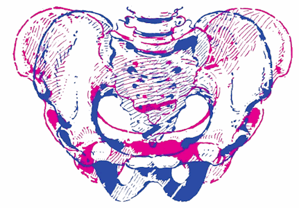 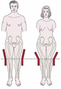
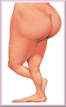
The wider pelvis of a woman results in the femur (upper leg bone) joining the hips to the knee at a different (less vertical) angle than in men; that’s why women walk slightly differently to men. Most notably women have a slightly greater swinging of the hips and hold their arms out a little more for balance and to keep space with their wider hips. The differences are very subtle but evolution has made us all ultra-sensitive at detecting male vs female characteristics, and a male rather than female gait can be a give-away at a distance. Some transwomen put a lot of time and effort in to "walking like a woman" but it's not a natural motion for their skeleton, and is difficult and even uncomfortable to sustain. Also, when consciously trying to trying to adopt a female gait, it's all too easy to exaggerate this and arrive at a 'cat walk model' or 'gay man' motion that actually attracts unwanted attention.
Long Bones and other
Bones
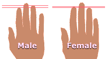
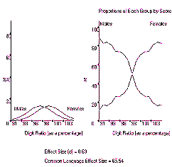
Finger
length ratio has become a widely accepted form of sex differentiation.
Most men have an index finger (digit 2, or 2D) shorter than their ring finger
(digit 4, or 4D), whilst most women have an index finger that is as long or longer
than their ring. finger. Statistically, women with a 2D to 4D length ratio of
0.98:1 are The difference in finger length seems to be due to the action of "male" androgen hormones on the skeleton of a developing foetus. This is confirmed by the fact that suffers of Androgen Insensitivity Syndrome (who are genetically XY male, but not affected by Androgen) have a statistically female digit ratio, whilst women who suffer from congenital adrenal hyperplasia (which raises their androgen levels before birth) have a statistically male ratio.
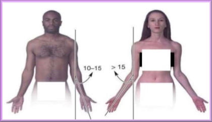
The bones in the arms of XY men are optimised for physical activities such as throwing, chopping and digging. Those of women are optimised for carrying young children and breast feeding a baby. As a result there is significant difference between male and female skeletons in what is called the "carrying angle" of the elbow. When the arms are held out at the side of the body but with the palms facing forward, a man's lower arm and hands typically point about 10 degrees away from the body, whilst those of a XX woman are at least 15 degrees.
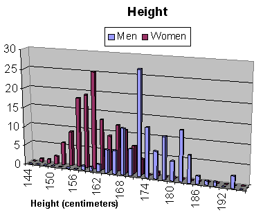
Height Height is a good indicator of sex, although a world-wide increase in height of several inches over the last 60 years due to better nutrition has confused the statistics and age-based percentiles. Future archaeology digs will face the problem that on average a man born in 1940 will be statistically similar in adult height to a woman born in 2000. The following table indicates some examples of the differentiation between average male and female heights in surveys taken since 2003. Surveys consistently show that the average cis-man is about 5.5 inches (14 cm) taller than the average cis-woman.
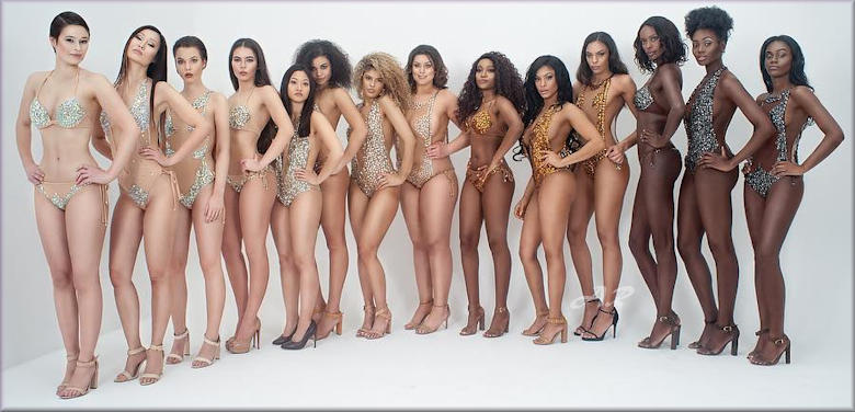
Trans Awareness Week. Although all are presumably genetically XY male, a huge diversity in height is obvious. Summary There is no definite 100% way of distinguishing a male from a female skeleton, but here is a list of the major criteria to be considered.
Surgical Feminisation of the Male Skeleton You may now want to read the article on this site about feminisation surgery on the genetic male.
Comparison Sites I used to include here a link to a website with an applet for comparing your height against male and female averages. At 5ft 9in I was boringly average as a man, but apparently only 3% of women were taller - which I admittedly found hard to believe given the number of young women that were towering over me every day! Unfortunately that website has gone off-line. If you can recommend a particularly good site for the comparison of male and female characteristics such as height, please send me an email: annie.richards@hotmail.com
|
|||||||||||||||||||||||||||||||||||||||||||||||||||||||||||||||||||||||||||||||||||||||||||||||||||||||||||||||||||||||||||||||||||||||||||||||||||||||||||||||||||||||||||||||||||||||||||||||||||||||||||||||||||||||||||||||||||||||||||||||||||||||||||||||||||||||||||||||||||||||||||||||||||
Last updated: 22 January, 2012


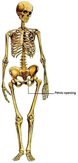

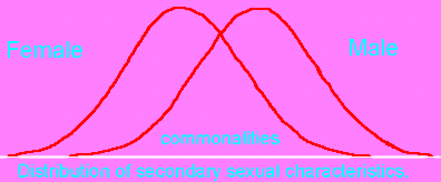 It is actually
quite difficult to distinguish between male and female skeletons as there
is a clear range of overlap between the sexes for many measures.
Indeed, there is in fact no certain, guaranteed, method of telling
the sex of a skeleton from its examination and inspection alone. For
example criminologists have misidentified the sex of murder victims,
whilst X-rays
of Egyption Pharaoh Tutankhamen failed to conclude with high
certainty if the skeleton was male or female.










 rare and anything less than that is strong indicator of a 'male' natal sex.




 Back to Articles
 (No spaces)